home | anatomy | physiology | pathology | clinical guides

1. Lungs position and relationship. left lung has 2 lobes and right 3 lobes.
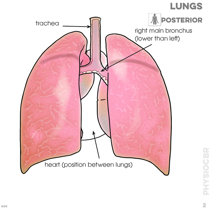
2. trachea; right branch of bronchus; position of heart
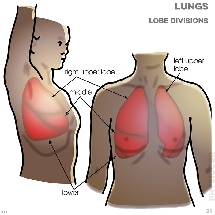
21. lungs, lobe divisions: Upper left lobe (ULL); upper right lobe; middle; lower; LLL
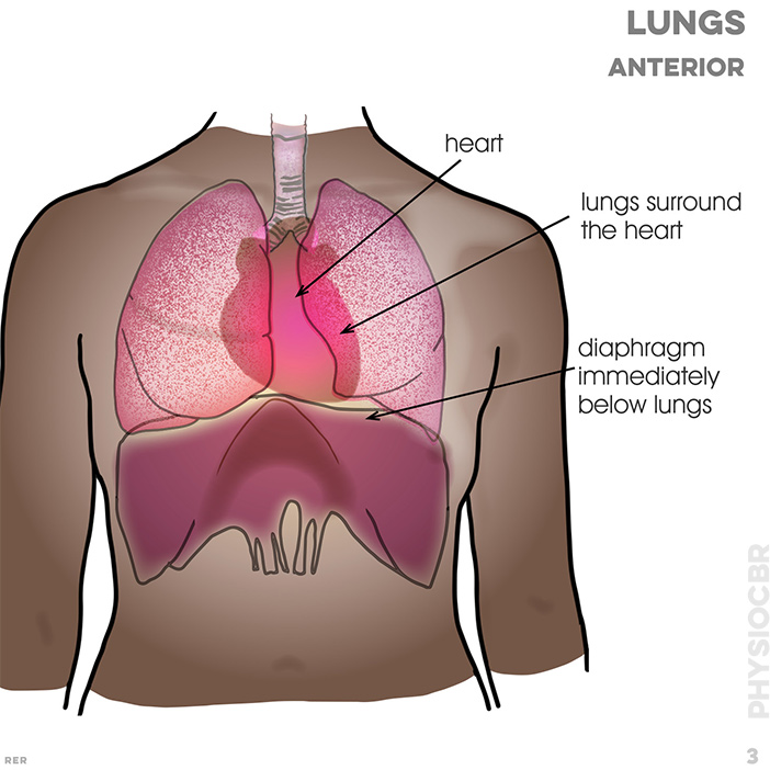
3. lungs surround the heart; diaphragm lies immediately under lungs

4. Rib cage (anterior/posterior): rib cage surrounds lungs; cartilage (includes sternum)
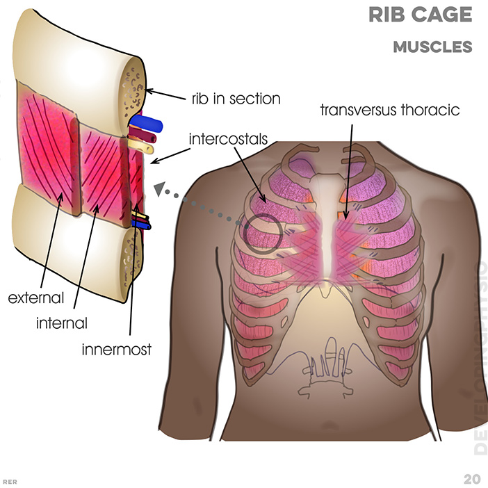
20. Rib cage and muscles: rib cage surrounds lungs; intercostal muscles: innermost, internal, external attach to ribs; transversus thoracic muscle; cartilage; diaphragm
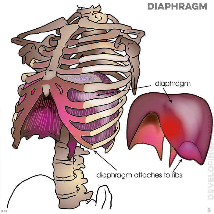
5. Diaphragm: diaphragm muscle attaches to ribs and spine
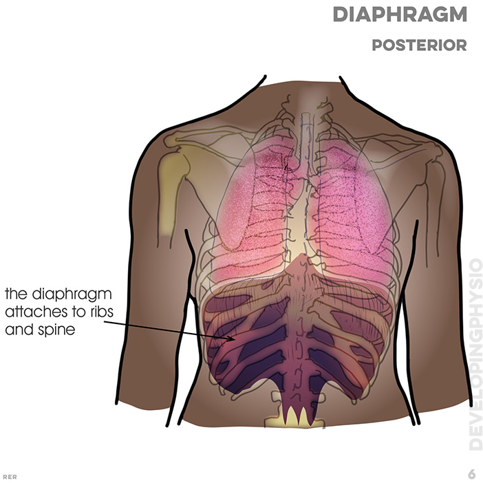
6. Diaphragm: posterior: diaphragm attaches to ribs and spine
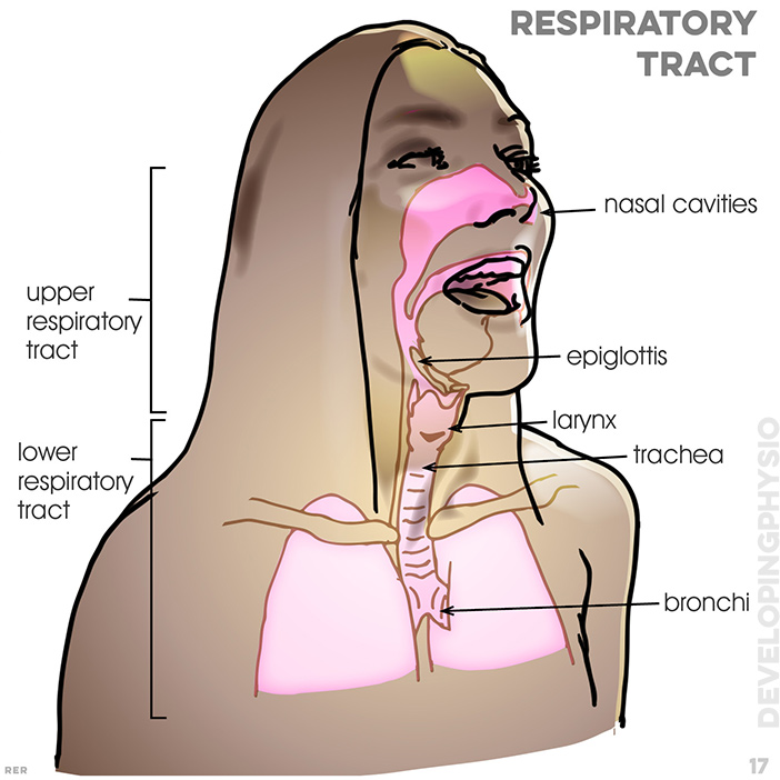
17. Respiaratory tract : nasal cavities; tongue; epiglottis; larynx; trachea; bronchi
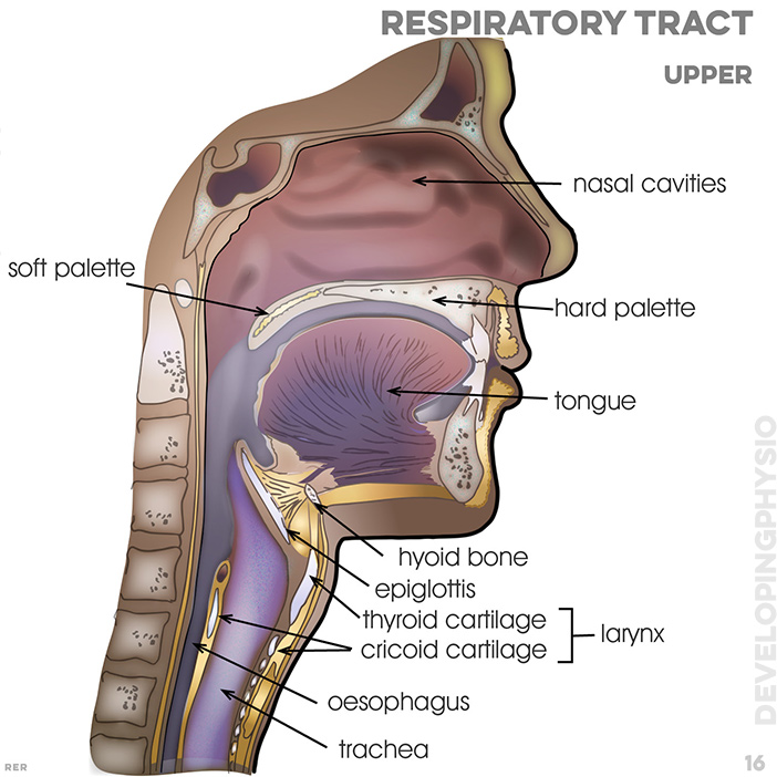
16. Respiratory tract (upper): nasal cavities; hard palette; soft palette; tongue; hyoid bone; epiglottis; thyroid cartilage and cricoid cartilage (make up larynx); oesophagus; trachea
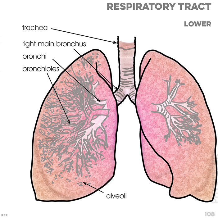
108. Lower respiratory tract : trachea, bronchus, bronchi, bronchioles, alveoli
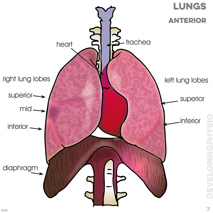
7. Lungs, anterior: trachea; heart; right lung lobes: superior, mid,inferior; left lung lobes: superior, inferior; diaphragm

101. Heart ventricles and valves: left ventricle; right ventricle; pulmonary valve, lung capillaries

106. Heart ventricles and valves: left ventricle; right ventricle; pulmonary valve, mitral valve; tricuspid valve, pulmonary arteries, left and right atrium
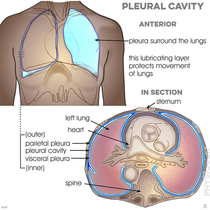
8. Pleural cavity: pleura surround the lungs; this lubricating layer protects movement of the lungs; in section: sternum, left lung, heart, parietal pleura, pleural cavity, visceral pleura, spine
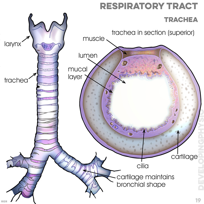
19. Respiratory tract, trachea: larynx; trachea; trachea in section (superior); muscle; lumen; mucal layer; cilia; cartilage; cartilage maintains bronchial shape
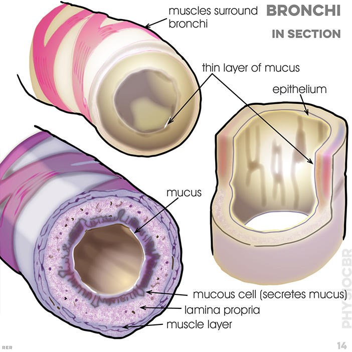
14. Bronchi in section: muscles surround bronchi; mucus; epithelium; mucous cell (secrete mucus); lamina propria; muscle layer
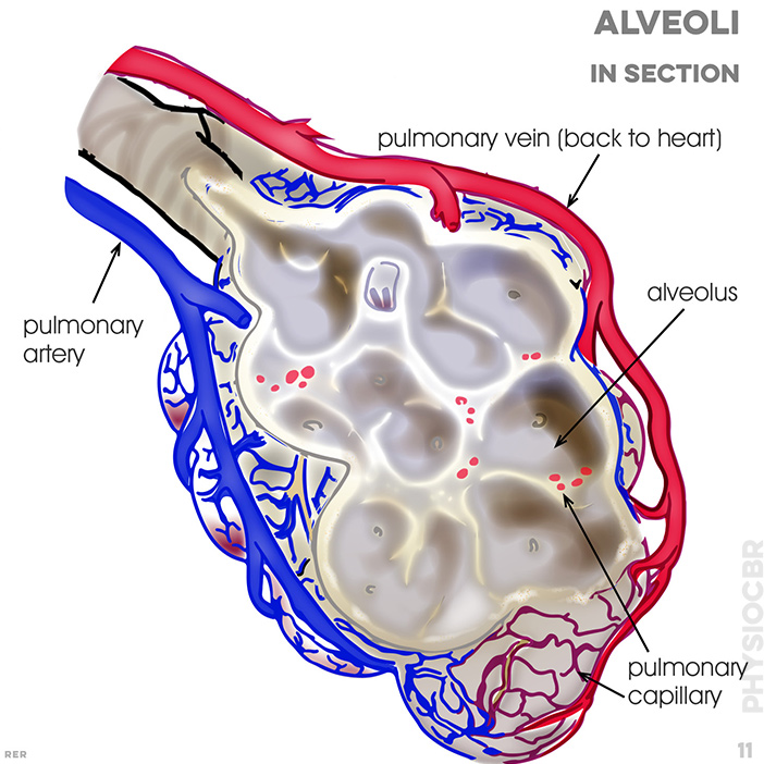
11. Alveoli in section: pulmonary vein; alveolar sac; pulmonary capillary; pulmonary artery
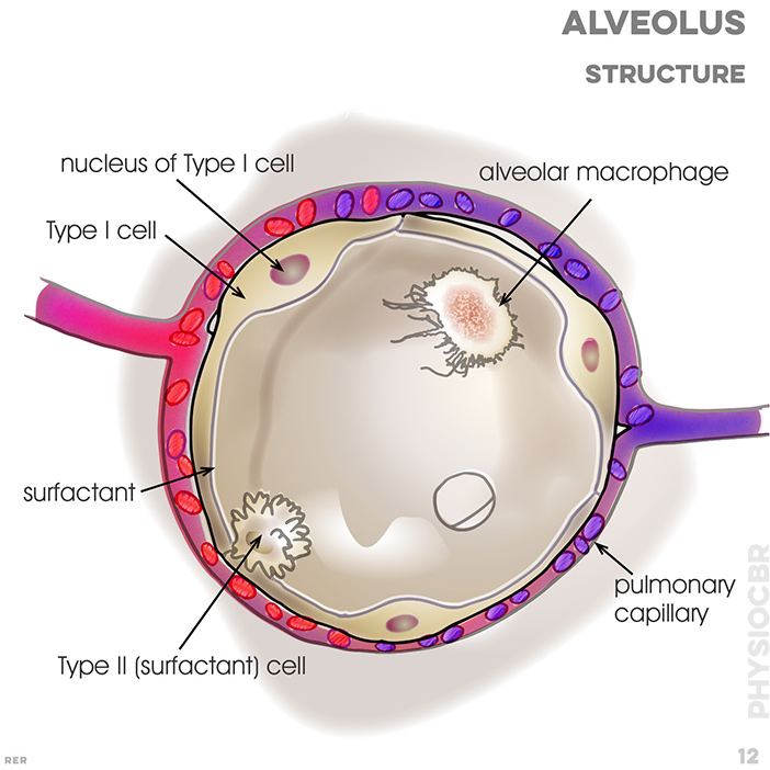
12. Alveolus structure: nucleus of Type I cell; alveolar macrophage; pulmonary capillary; type I cell; type II (surfactant) cell
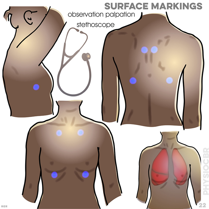
22. Surface markings, stethoscope points
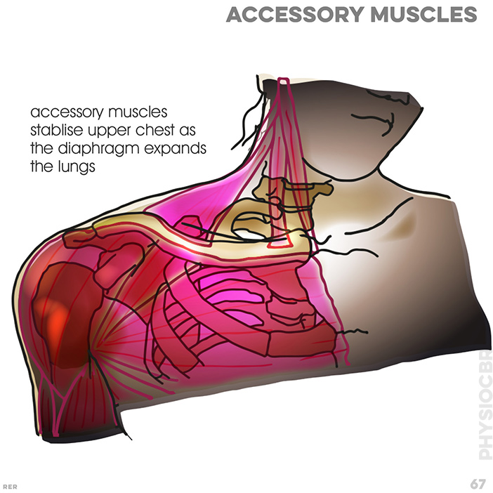
67. shoulder and chest muscles are accessory to helping lung movement
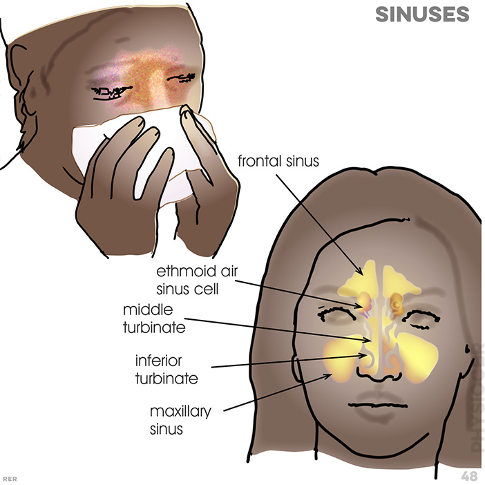
48. sinuses; frontal, ethmoid air, middle turbinate, inferiro turbinate, maxillary
home | anatomy | physiology | pathology | clinical guides

![]() Quick quiz?
on anatomy
Quick quiz?
on anatomy
![]() Feedback
memo while you remember it!
Feedback
memo while you remember it!
The key regions: RIBS: (and heart position); LUNGS: position within ribs and around heart (note left lung different to right); DIAPHRAGM: immediately beneath lungs with edges attached to lowest ribs; PLEURA: surrounds the lungs in protective function; RIB MUSCLES: between each rib to aid breathing; BRONCHI: and respiratory tract; ALVEOLI: where diffusion occurs;